Shoulder Tendon And Ligament Anatomy / shoulder tendons anatomy - Google Search | Human anatomy ... - Other smaller muscles and tendons surround the knee joint as well.. Both tendons and ligaments are dense regular connective tissue, because of its two properties: Shoulder anatomy is an elegant piece of machinery having the greatest range of motion of any joint in the body. Learn about their differences and the common injuries that affect them here. These ligaments are main source of stability for the shoulder. Learn vocabulary, terms and more with flashcards, games and other study tools.
The patellar tendon on the front of the knee is part of the quadriceps mechanism. Have ligaments shoulder joint pain? These tendinous insertions along with the articular capsule, the coracohumeral ligament, and the glenohumeral ligament complex subscapular bursa is located between the subscapularis tendon and the scapula. (3) a syndesmosis is a joint in which a ligament connects two bones, allowing for a little movement (amphiarthroses). In addition to the bones and joints, the shoulder contains a network of soft tissues, such as muscles, tendons, and ligaments.
Shoulder Muscles Diagram - Labeled Anatomy Chart Of Neck ... from o.quizlet.com Start studying shoulder ligaments and tendons. Learn vocabulary, terms and more with flashcards, games and other study tools. Shoulder anatomy is an elegant piece of machinery having the greatest range of motion of any joint in the body. However, many tendon and ligament injuries can be avoided through proper conditioning and training regimens and by not pushing a horse beyond its limits in racing or other competitions. Both tendons and ligaments are dense regular connective tissue, because of its two properties: Tendon and ligament injuries often go hand in hand with horses involved in vigorous athletic pursuits. In its complex function, the shoulder must be. The distal joint between the tibia and fibula is an example of a.
Tendons and ligaments are bands of connective tissue that help stabilize the body and allow movement.
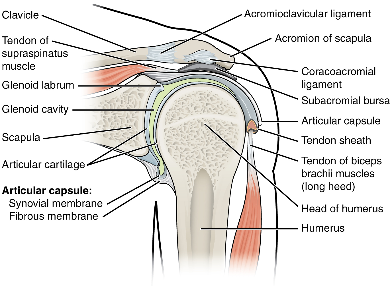
Start studying shoulder ligaments and tendons. Contents of ri = long head of biceps tendon, superior glenohumeral ligament, glenohumeral capsule. Original editors mary harris , tom lawlor , patrick bales , misty hillin , rick wetherald as part of the texas evidence based practice project. The ligaments of the shoulder joint in the shoulder joint, the majority of the ligaments are thickenings of the joint capsule. Shoulder muscles and shoulder tendons. Both tendons and ligaments are dense regular connective tissue, because of its two properties: Joints can be grouped by their structure into fibrous, cartilaginous, and synovial joints. The shoulder joint (glenohumeral joint) is a ball and socket joint between the scapula and the humerus. Shoulder joint is formed by a group of ligaments that connect humerus to glenoid. Anatomy of the canine shoulder (scapula, humerus, ligaments, shoulder joint, muscles and tendons) on ct. Links the coracoid to the acromium and forms the. In addition to the bones and joints, the shoulder contains a network of soft tissues, such as muscles, tendons, and ligaments. Tendon and ligament injuries often go hand in hand with horses involved in vigorous athletic pursuits.
It usually results from your tendon being pinched by. Original editors mary harris , tom lawlor , patrick bales , misty hillin , rick wetherald as part of the texas evidence based practice project. Amyn rajani is the best orthopaedic doctor in south, central and suburban mumbai to treat all shoulder disorders. Muscles allow us to move by pulling on bones. Tendons and ligaments are bands of connective tissue that help stabilize the body and allow movement.
9.6 Anatomy of Selected Synovial Joints - Anatomy and ... from opentextbc.ca Once stretched, they tend to stay. The distal joint between the tibia and fibula is an example of a. Shoulder muscles and shoulder tendons. (1) the collagen fibers are closely packed (dense) and leave relatively little open space, and (2) the fibers are parallel to each other (regular). Tendon and ligament injuries often go hand in hand with horses involved in vigorous athletic pursuits. The patellar tendon on the front of the knee is part of the quadriceps mechanism. The muscles involved in the anatomy of the shoulder are many, with each contributing to the vast range of motion and stability. The shoulder joint (glenohumeral joint) is a ball and socket joint between the scapula and the humerus.
Anatomy of the canine shoulder (scapula, humerus, ligaments, shoulder joint, muscles and tendons) on ct.
The brachial plexus anatomy animation: However, many tendon and ligament injuries can be avoided through proper conditioning and training regimens and by not pushing a horse beyond its limits in racing or other competitions. Many muscles, tendons, ligaments and cartilage form the soft tissue components of the shoulder's anatomy. Start studying shoulder ligaments and tendons. The ca ligament along with the acromial process create the outlet of the shoulder thru which passes the supraspinatus tendon of the rotator cuff. Shoulder muscles and shoulder tendons. The patellar tendon on the front of the knee is part of the quadriceps mechanism. Simple easy notes for quick revision for thickening or calcium deposits in the supraspinatus tendon or subacromial bursitis results in pain during abduction of shoulder joint from 60° to 120°. Amyn rajani is the best orthopaedic doctor in south, central and suburban mumbai to treat all shoulder disorders. The distal joint between the tibia and fibula is an example of a. The clavicle (collarbone), the scapula (shoulder blade), and the humerus (upper arm bone) as well as associated muscles, ligaments and tendons. In addition to the bones and joints, the shoulder contains a network of soft tissues, such as muscles, tendons, and ligaments. A joint capsule is a watertight sac that surrounds a joint.
In addition to the bones and joints, the shoulder contains a network of soft tissues, such as muscles, tendons, and ligaments. (3) a syndesmosis is a joint in which a ligament connects two bones, allowing for a little movement (amphiarthroses). The shoulder joint (glenohumeral joint) is a ball and socket joint between the scapula and the humerus. (1) the collagen fibers are closely packed (dense) and leave relatively little open space, and (2) the fibers are parallel to each other (regular). Injury of tendons and ligaments remodel with scar formation with differences in themselves.
PPT - The Musculoskeletal Examination in the Elderly ... from image.slideserve.com Anteriorly the subscapularis tendon is separated from the supraspinatus tendon by a gap, the rotator interval another important ligament, the coracoacromial ligament (cal). Roots, trunks, divisions, cords, branches, clinical anatomy. Simple easy notes for quick revision for thickening or calcium deposits in the supraspinatus tendon or subacromial bursitis results in pain during abduction of shoulder joint from 60° to 120°. The patellar tendon on the front of the knee is part of the quadriceps mechanism. The difference between ligaments and tendon. Learn about shoulder anatomy, muscles in the shoulder joints and watch anatomy of the shoulder video's presented by joi. (3) a syndesmosis is a joint in which a ligament connects two bones, allowing for a little movement (amphiarthroses). This instability is countered by the strength of the rotator cuff muscles, tendons, ligaments, and the glenoid labrum.
In addition to the bones and joints, the shoulder contains a network of soft tissues, such as muscles, tendons, and ligaments.
Although scarring depends on the quality and quantity of the injured tissues, it can be. Start studying shoulder ligaments and tendons. (3) a syndesmosis is a joint in which a ligament connects two bones, allowing for a little movement (amphiarthroses). More about dental anatomy and periodontal ligaments you can find in the article about the anatomy of the teeth and this interesting video tutorial. Injury of tendons and ligaments remodel with scar formation with differences in themselves. The shoulder joint (glenohumeral joint) is a ball and socket joint between the scapula and the humerus. The anatomy of the provides the strength and functionality of the upper body. Tendons and ligaments commonly sustain injuries, which usually have similar symptoms and treatments. Dr.g bhanu prakash animated medical videos. Original editors mary harris , tom lawlor , patrick bales , misty hillin , rick wetherald as part of the texas evidence based practice project. (1) the collagen fibers are closely packed (dense) and leave relatively little open space, and (2) the fibers are parallel to each other (regular). Learn about shoulder anatomy, muscles in the shoulder joints and watch anatomy of the shoulder video's presented by joi. Superior glenohumeral ligament and coracohumeral ligament are the primary restraints to posterior acromioclavicular ligament anatomy.
Other smaller muscles and tendons surround the knee joint as well shoulder tendon anatomy. Tendons and ligaments are bands of connective tissue that help stabilize the body and allow movement.